
Guide
Understanding your newborn’s ear shape
Every baby’s ears are a little different. Knowing the names of the main parts makes it easier to describe what you see and to follow what a clinician explains.
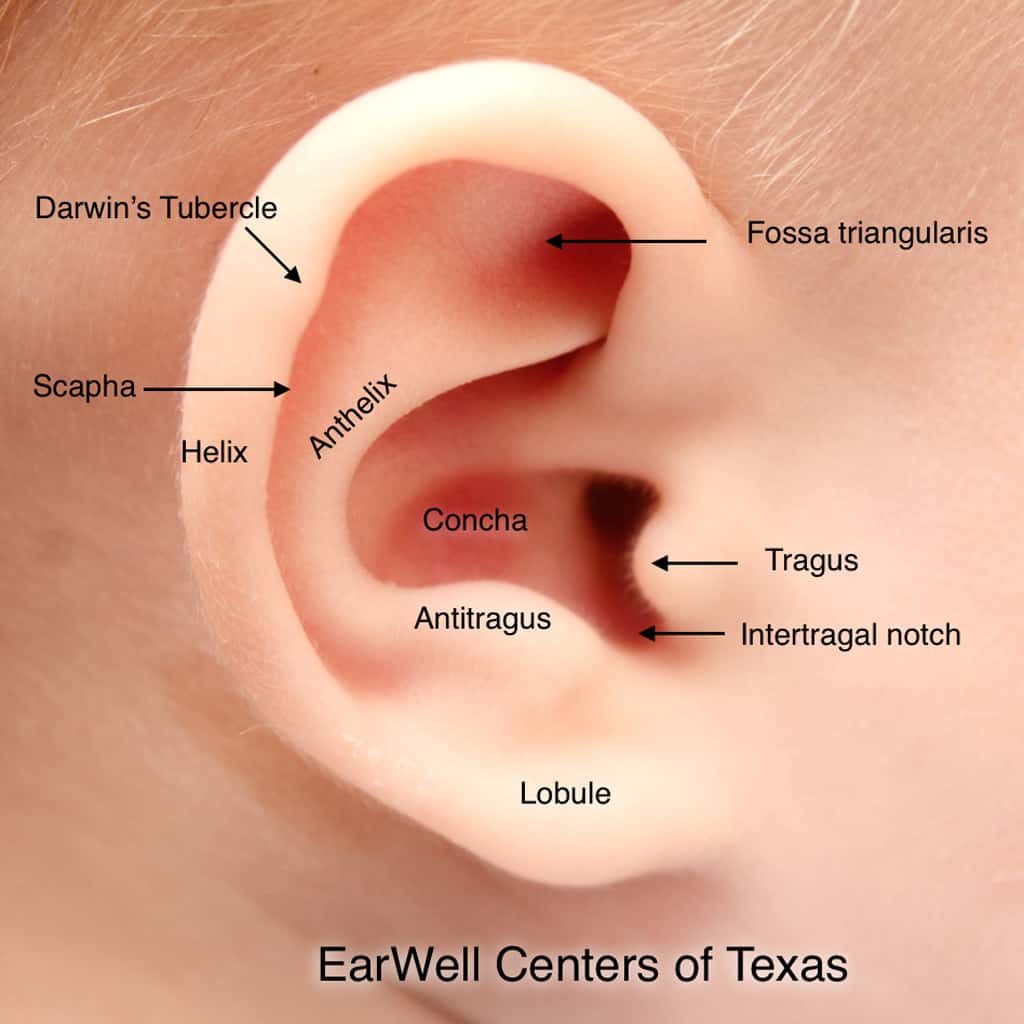
The parts of the outer ear
- Helix: the curled outer rim of the ear.
- Antihelix: the curved ridge inside the rim. Near the top it splits into two branches.
- Scapha: the groove between the helix and antihelix.
- Fossa triangularis: the small hollow between the two upper branches of the antihelix.
- Concha: the bowl of the ear leading to the ear canal.
- Tragus and antitragus: the small bumps in front of and below the ear canal.
- Lobule: the earlobe.
How much variation is normal?
Ears vary in size, angle and the depth of their folds, and many babies’ two ears are not identical. Some newborn ears look squashed or folded for a few days after birth and then relax. A difference becomes worth checking when a fold, ridge, tightness or angle stands out or persists.
What causes ear shape differences?
- Position before birth. Pressure in the womb or during birth can fold a soft ear.
- Development of the ear’s cartilage and muscles. Small differences in how the ear formed can create an extra fold or a missing curve.
- Family traits. Some ear shapes run in families.
In most cases there is no single cause, and it is not something parents did. When tissue is missing, as in microtia, the cause is different and a broader assessment is recommended.
When to ask for an assessment
Ask if a fold, ridge, point, tight rim or prominent angle is noticeable, if it has not changed after a few days, or if one ear looks smaller or incomplete. You do not need to be sure before you call. See common ear conditions.
Questions about newborn ear shape
What are the main parts of the ear?
The helix (outer rim), antihelix (inner ridge), scapha (groove between them), concha (bowl), tragus, antitragus and lobule (earlobe).
Is it normal for my baby’s ears to look different from each other?
Some difference is common. A fold, ridge, point or angle that stands out or does not change after a few days is worth having checked.
What causes newborn ear shape differences?
Position before birth, how the ear’s cartilage developed and family traits can all play a part. Usually there is no single cause.
When should I ask a clinician to look at my baby’s ears?
If something stands out, persists after a few days, or one ear looks smaller or incomplete. Call 214-540-1428 for an evaluation.
Questions about your baby’s ears?
Request an evaluation with EarWell Centers or call our team. We will help you understand the next step.